
|
Abhishek Singh Sambyal
email (academics):
(enable JavaScript to reveal)
I am a Postdoctoral Researcher in the Intelligent Medical Systems (IMEDS) Group at the University of Oulu, working with Prof. Aleksei Tiulpin. My research interests lie in uncertainty quantification, confidence calibration and reliability of deep neural networks for medical imaging applications. Previously, I completed my PhD in the Department of Computer Science and Engineering at the Indian Institute of Technology Ropar, India, where I was advised by Dr. Deepti R. Bathula and Dr. Narayanan C. Krishnan. |
Recent News
|
Publications |
|
|
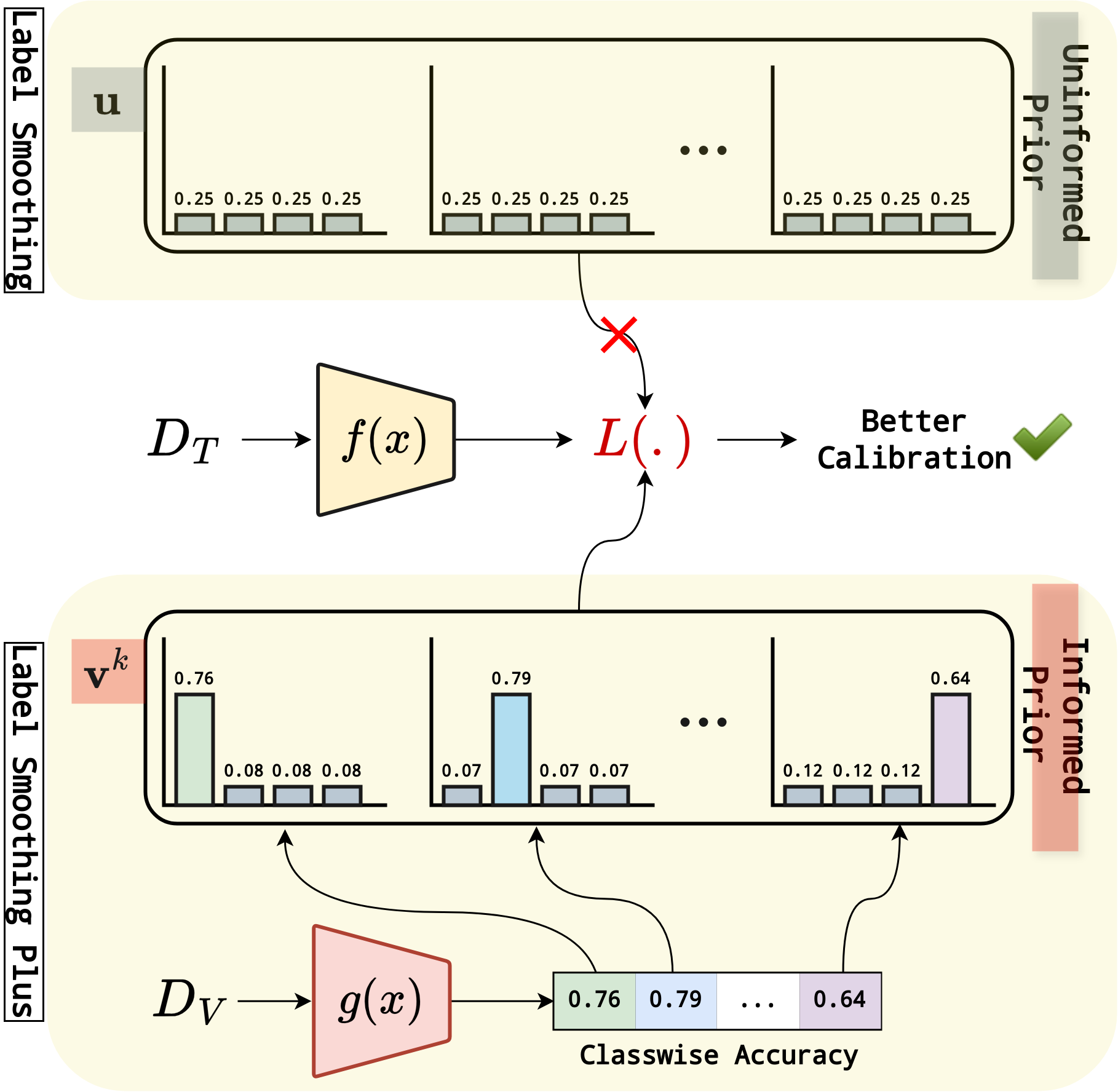
LS+: Informed Label Smoothing for Improving Calibration in Medical Image ClassificationMICCAI RISE Registration Award
paper
supplementary
code
Deep Neural Networks (DNNs) exhibit exceptional performance in various tasks; however, their susceptibility to miscalibration poses challenges in healthcare applications, impacting reliability and trustworthiness. Label smoothing, which prefers soft targets based on uniform distribution over labels, is a widely used strategy to improve model calibration. We propose an improved strategy, Label Smoothing Plus (LS+), which uses class-specific prior that is estimated from validation set to account for current model calibration level. We evaluate the effectiveness of our approach by comparing it with state-of-the-art methods on three benchmark medical imaging datasets, using two different architectures and several performance and calibration metrics for the classification task. Experimental results show notable reduction in calibration error metrics with nominal improvement in performance compared to other approaches, suggesting that our proposed method provides more reliable prediction probabilities.
@inproceedings{lsplus,
  author    = {Sambyal, Abhishek Singh and
               Niyaz, Usma and
               Shrivastava, Saksham and
               Krishnan, Narayanan C. and
               Bathula, Deepti R.},
  title     = {{LS+}: Informed Label Smoothing for Improving Calibration in
               Medical Image Classification},
  booktitle = {Medical Image Computing and Computer Assisted Intervention --
               MICCAI 2024},
  pages     = {513--523},
  year      = {2024}
}
|
|
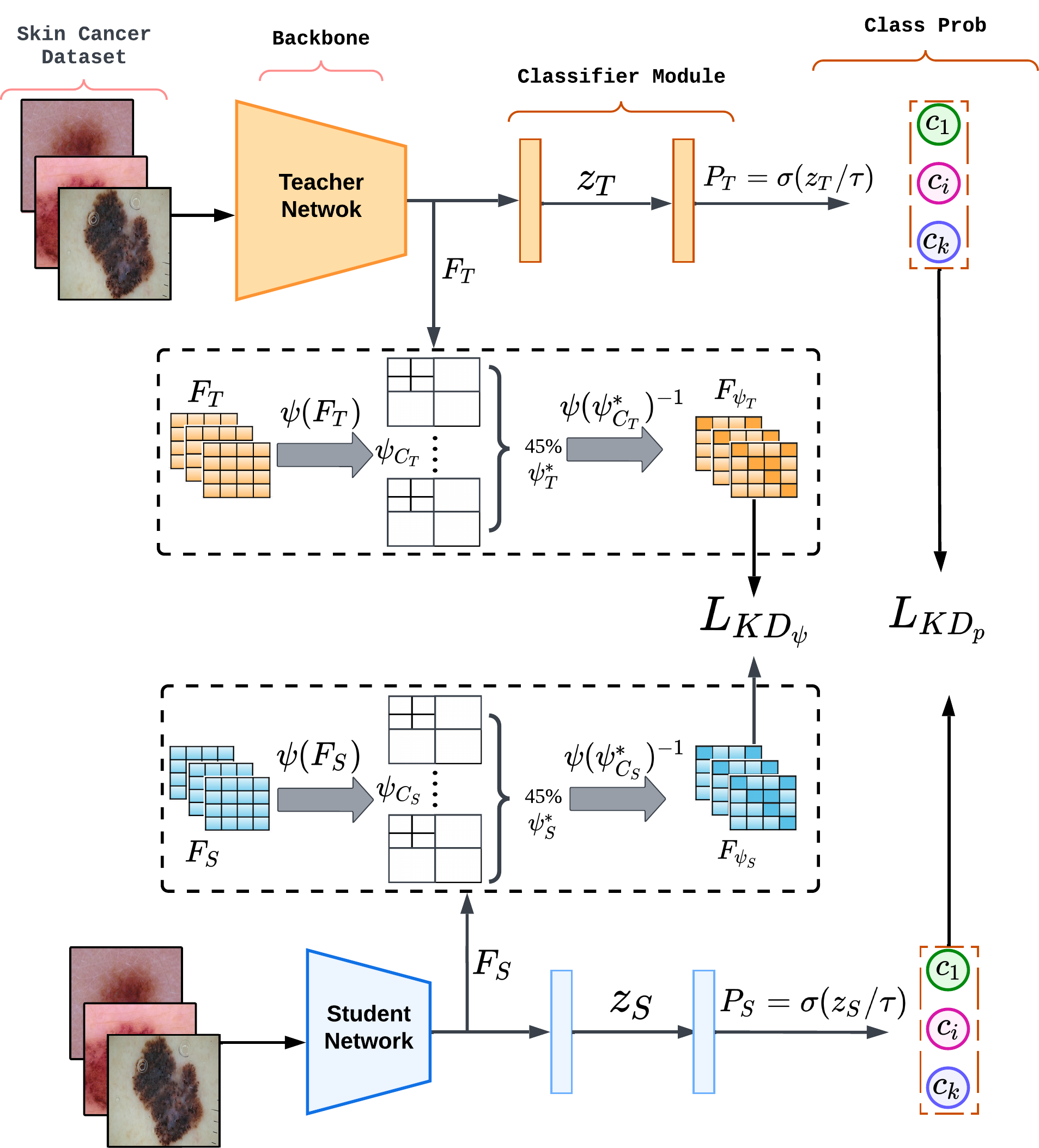
Wavelet-Based Feature Compression for Improved Knowledge DistillationISBI Travel Award
paper
code
Deep learning (DL) models can achieve state-of-the-art performance but at the cost of high computation and memory requirements. Due to their large capacity, DL models have the tendency to learn redundant features. In this work, we exploit this redundancy to improve model compression. Knowledge distillation (KD) aims to achieve model compression by transferring knowledge from a large, complex model to a simple, lightweight model. We propose an enhanced KD strategy that improves the efficiency of the distillation process by compressing the feature maps using Discrete Wavelet Transformation (DWT), which helps capture crucial features from complex biomedical signals. Retaining and sharing only the most informative and discriminating features facilitates more effective feature mimicking. Extensive experiments using two benchmark datasets for Melanoma and Histopathology image classification tasks demonstrate the superiority of our proposed method over existing techniques. We further establish the generalizability and robustness of our method using two different teacher-student architectures and ablation studies.
@inproceedings{usma-waveletkd-isbi2024,
  author    = {Niyaz, Usma and
               Sambyal, Abhishek Singh and
               Bathula, Deepti R.},
  title     = {Wavelet-Based Feature Compression for Improved Knowledge
               Distillation},
  booktitle = {2024 IEEE International Symposium on Biomedical Imaging (ISBI)},
  pages     = {1--4},
  year      = {2024},
  doi       = {10.1109/ISBI56570.2024.10635879}
}
|
|
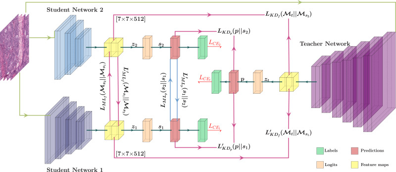
Leveraging Different Learning Styles for Improved Knowledge Distillation in Biomedical Imaging
journal
arxiv
Learning style refers to a type of training mechanism adopted by an individual to gain new knowledge. As suggested by the VARK model, humans have different learning preferences, like Visual (V), Auditory (A), Read/Write (R), and Kinesthetic (K), for acquiring and effectively processing information. Our work endeavors to leverage this concept of knowledge diversification to improve the performance of model compression techniques like Knowledge Distillation (KD) and Mutual Learning (ML). Consequently, we use a single-teacher and two-student network in a unified framework that not only allows for the transfer of knowledge from teacher to students (KD) but also encourages collaborative learning between students (ML). Unlike the conventional approach, where the teacher shares the same knowledge in the form of predictions or feature representations with the student network, our proposed approach employs a more diversified strategy by training one student with predictions and the other with feature maps from the teacher. We further extend this knowledge diversification by facilitating the exchange of predictions and feature maps between the two student networks, enriching their learning experiences. We have conducted comprehensive experiments with three benchmark datasets for both classification and segmentation tasks using two different network architecture combinations. These experimental results demonstrate that knowledge diversification in a combined KD and ML framework outperforms conventional KD or ML techniques (with similar network configuration) that only use predictions with an average improvement of 2%. Furthermore, consistent improvement in performance across different tasks, with various network architectures, and over state-of-the-art techniques establishes the robustness and generalizability of the proposed model.
@article{NIYAZ-cbm-2023,
  author  = {Niyaz, Usma and
             Sambyal, Abhishek Singh and
             Bathula, Deepti R.},
  title   = {Leveraging Different Learning Styles for Improved Knowledge
             Distillation in Biomedical Imaging},
  journal = {Computers in Biology and Medicine},
  volume  = {168},
  pages   = {107764},
  year    = {2024},
  issn    = {0010-4825},
  doi     = {10.1016/j.compbiomed.2023.107764}
}
|
|
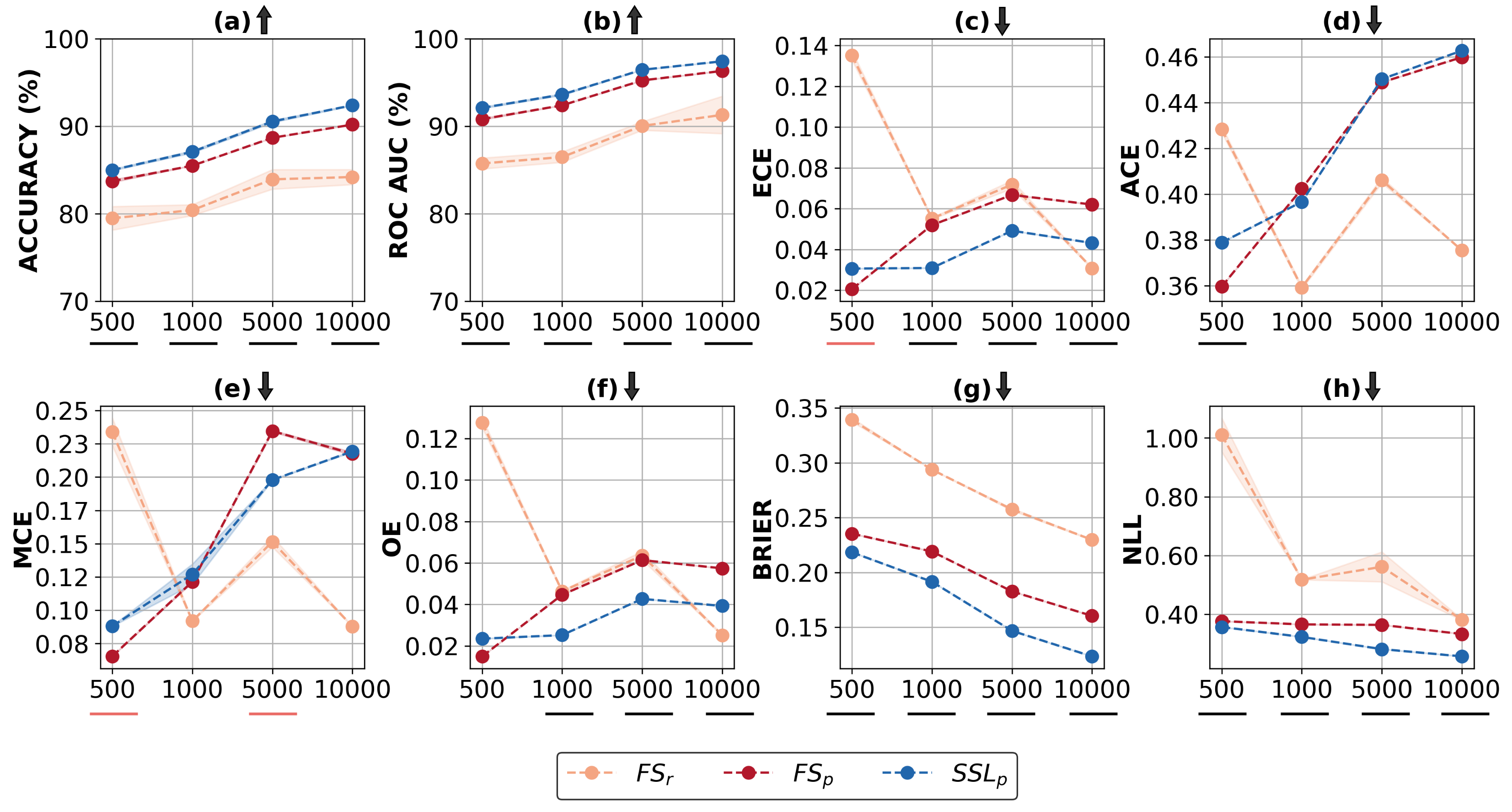
Understanding Calibration of Deep Neural Networks for Medical Image Classification
journal
arxiv
In the field of medical image analysis, achieving high accuracy is not enough; ensuring well-calibrated predictions is also crucial. Confidence scores of a deep neural network play a pivotal role in explainability by providing insights into the model's certainty, identifying cases that require attention, and establishing trust in its predictions. Consequently, the significance of a well-calibrated model becomes paramount in the medical imaging domain, where accurate and reliable predictions are of utmost importance. While there has been a significant effort towards training modern deep neural networks to achieve high accuracy on medical imaging tasks, model calibration and factors that affect it remain under-explored. To address this, we conducted a comprehensive empirical study that explores model performance and calibration under different training regimes. We considered fully supervised training, which is the prevailing approach in the community, as well as rotation-based self-supervised method with and without transfer learning, across various datasets and architecture sizes. Multiple calibration metrics were employed to gain a holistic understanding of model calibration. Our study reveals that factors such as weight distributions and the similarity of learned representations correlate with the calibration trends observed in the models. Notably, models trained using rotation-based self-supervised pretrained regime exhibit significantly better calibration while achieving comparable or even superior performance compared to fully supervised models across different medical imaging datasets. These findings shed light on the importance of model calibration in medical image analysis and highlight the benefits of incorporating self-supervised learning approach to improve both performance and calibration.
@article{understandingcalibration-2023,
  author  = {Sambyal, Abhishek Singh and
             Niyaz, Usma and
             Krishnan, Narayanan C. and
             Bathula, Deepti R.},
  title   = {Understanding Calibration of Deep Neural Networks for Medical
             Image Classification},
  journal = {Computer Methods and Programs in Biomedicine},
  volume  = {242},
  pages   = {107816},
  year    = {2023},
  issn    = {0169-2607},
  doi     = {10.1016/j.cmpb.2023.107816}
}
|
|
Towards Reducing Aleatoric Uncertainty for Medical Imaging Tasks
presentation
poster
arxiv
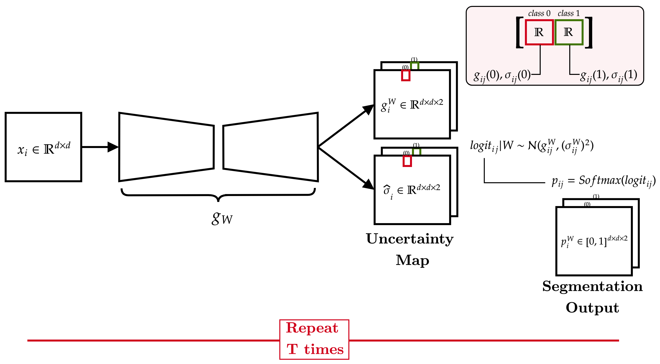
In safety-critical applications like medical diagnosis, certainty associated with a model's prediction is just as important as its accuracy. Consequently, uncertainty estimation and reduction play a crucial role. Uncertainty in predictions can be attributed to noise or randomness in data (aleatoric) and incorrect model inferences (epistemic). While model uncertainty can be reduced with more data or bigger models, aleatoric uncertainty is more intricate. This work proposes a novel approach that interprets data uncertainty estimated from a self-supervised task as noise inherent to the data and utilizes it to reduce aleatoric uncertainty in another task related to the same dataset via data augmentation. The proposed method was evaluated on a benchmark medical imaging dataset with image reconstruction as the self-supervised task and segmentation as the image analysis task. Our findings demonstrate the effectiveness of the proposed approach in significantly reducing the aleatoric uncertainty in the image segmentation task while achieving better or on-par performance compared to the standard augmentation techniques.
@inproceedings{trau,
  author    = {Sambyal, Abhishek Singh and
               Krishnan, Narayanan C. and
               Bathula, Deepti R.},
  title     = {Towards Reducing Aleatoric Uncertainty for Medical Imaging
               Tasks},
  booktitle = {2022 IEEE 19th International Symposium on Biomedical Imaging
               (ISBI)},
  pages     = {1--4},
  year      = {2022},
  doi       = {10.1109/ISBI52829.2022.9761638}
}
|
|
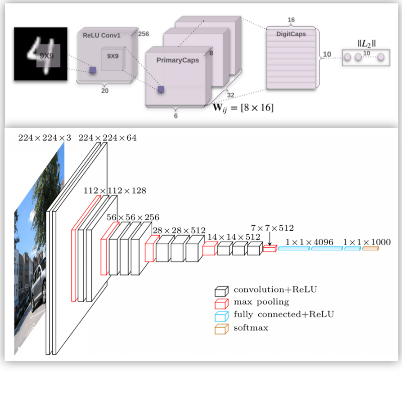
Advances in Deep Learning Techniques for Medical Image AnalysisDeep learning is contributing to the high level of services to the healthcare sector. As the digital medical data is increasing exponentially with time, early detection and prediction of diseases are becoming more efficient because of the deep learning techniques which reduce the fatality rate to a great extent. The main focus of this paper is to provide the comprehensive review of deep learning in the domain of medical image processing and analysis. We have demonstrated the use of new deep learning architectures in oncology for the prediction of different types of cancer like the brain, lung, skin, etc. The state-of-the-art architectures effectively carry out analysis of 2D and 3D medical images to make the diagnosis of patients faster and more accurate. The use of popular approaches in machine learning such as ensemble and transfer learning with fine-tuning of parameters improve the performance of the deep neural networks in medical image analysis. The existing deep networks urge the new image classification network called Capsule Network (CapsNet) to make the classification and detection comparatively better. The equivariance characteristics of CapsNet make it more influential as it discourages the effect of any structural invariance of an input image on the network.
@inproceedings{adlt,
  author    = {Niyaz, Usma and
               Sambyal, Abhishek Singh and
               Padha, Devanand},
  title     = {Advances in Deep Learning Techniques for Medical Image Analysis},
  booktitle = {2018 Fifth International Conference on Parallel, Distributed
               and Grid Computing (PDGC)},
  year      = {2018},
  doi       = {10.1109/PDGC.2018.8745790}
}
|
|
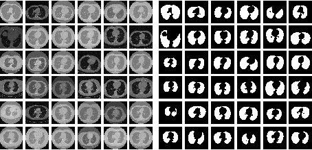
Evaluation of Deep Learning Model with Optimizing and Satisficing Metrics for Lung SegmentationThe segmentation in medical image analysis is a crucial and prerequisite process during the diagnosis of the diseases. The need for segmentation is important to attain the region of interest where the probability of occurrence of an abnormality such as a nodule in the lungs or tumor in the brain is high. In this paper, we have proposed a new architecture called FS-Net which is a convolutional neural network-based model for the segmentation of lungs in CT scan images. It performs encoding of images into the feature maps and then decodes the feature maps into their respective lung masks. We have also trained the state-of-the-art U-Net on the same dataset and compared the results on the basis of optimizing and satisficing metrics. These metrics are useful for the selection of a better model with the maximum score at the satisfying condition. The FS-Net is computationally very efficient and achieves promising dice coefficient and loss score when compared with the U-Net taking one-third of the time.
@inproceedings{osm,
  author    = {Niyaz, Usma and
               Sambyal, Abhishek Singh and
               Padha, Devanand},
  title     = {Evaluation of Deep Learning Model with Optimizing and
               Satisficing Metrics for Lung Segmentation},
  booktitle = {Soft Computing for Problem Solving},
  year      = {2020}
}
|
|
Knowledge Abstraction from Textural Features of Brain MRI Images for Diagnosing Brain Tumor Using Statistical Techniques and Associative Classification
code
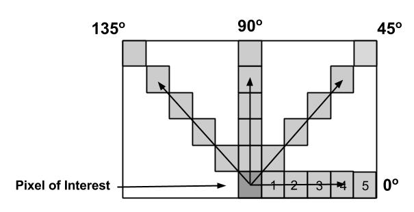
This paper presents a methodology for finding the association rules using associative classification which can be used to abstract knowledge from brain MRI images. Reducing the size of images using different thresholds help to reduce the complexity of the proposed system without affecting the correctness of these images. Textural features are taken into consideration because when there is a wide variation of features of discrete gray tone, the texture dominates more. Gray-Tone Spatial-Dependence matrices are calculated from images in which textural information is contained. The system uses a supervised learning approach for selecting the important features from different textural features. Using associative classification, the rules are generated from selected textural features which abstract the knowledge from the images.
@inproceedings{knowabs,
  author    = {Sambyal, Abhishek Singh and
               Asha, T.},
  title     = {Knowledge Abstraction from Textural Features of Brain {MRI}
               Images for Diagnosing Brain Tumor Using Statistical Techniques
               and Associative Classification},
  booktitle = {2016 International Conference on Systems in Medicine and
               Biology (ICSMB)},
  year      = {2016},
  doi       = {10.1109/ICSMB.2016.7915086}
}
|
Academic Service
|
Teaching
|